Home
Uncategories
Medial Epicondyle Elbow Xray : Elbow Radiology Key - A wide variety of medial lateral epicondyle options are available to there are 16 suppliers who sells medial lateral epicondyle on alibaba.com, mainly located in asia.
Medial Epicondyle Elbow Xray : Elbow Radiology Key - A wide variety of medial lateral epicondyle options are available to there are 16 suppliers who sells medial lateral epicondyle on alibaba.com, mainly located in asia.
Medial Epicondyle Elbow Xray : Elbow Radiology Key - A wide variety of medial lateral epicondyle options are available to there are 16 suppliers who sells medial lateral epicondyle on alibaba.com, mainly located in asia.. A wide variety of medial lateral epicondyle options are available to there are 16 suppliers who sells medial lateral epicondyle on alibaba.com, mainly located in asia. Elbow injuries often have characteristic radiological appearances, which may only be detected by the presence of soft. Posterolateral elbow dislocation, which may spontaneously. It develops where tendons in the forearm muscle connect to the bony part on the inside of the elbow. Incarcerated medial epicondyle fractures in association with elbow trauma are rare and an absolute indication for intervention.
The assessment of the elbow can be difficult because of the changing anatomy of the on the medial side the valgus force can lead to avulsion of the medial epicondyle. A condition characterized by pain in or near the lateral humeral epicondyle or in the forearm extensor muscle mass as a result of unusual strain. Elbow injuries often have characteristic radiological appearances, which may only be detected by the presence of soft. Incarcerated medial epicondyle fractures in association with elbow trauma are rare and an absolute indication for intervention. Radial neck, medial epicondyle or olecranon tip.
The Radiology Assistant Fractures In Children from radiologyassistant.nl 813 medial lateral epicondyle products are offered for sale by suppliers on alibaba.com. The medial epicondyle is the bony origin for the wrist flexors and involve the. Due to injury or irritation, they can become swollen and painful. Years at ossification (appear on xray) (1). Tennis elbow management a multimodal management. Related online courses on physioplus. Usually negative (evaluates more for differential diagnosis). Specifically in passive flexion of the elbow, it is subcutaneous and generally noticeable.
Medial epicondylitis is caused by any activity that places a valgus force on the elbow or that involves forcefully flexing the volar forearm muscles, as occurs during pitching.
Sometimes the medial epicondyl becomes trapped within the joint. However, several other sports and activities besides sports can also put you at risk. A fracture of the medial epicondyle of the elbow that is the third most common fracture seen in children and is usually seen in boys between the age of 9 and 14. The medial and lateral epicondyles are small bony tuberosities on the distal end of the humerus (fig. The top countries of suppliers are china, taiwan. The medial epicondyle is the bony origin for the wrist flexors and involve the. The assessment of the elbow can be difficult because of the changing anatomy of the on the medial side the valgus force can lead to avulsion of the medial epicondyle. Related online courses on physioplus. Performed with the elbow extended. Pain upon resisted wrist flexion. Bones visiable are he lateral and medial epicondyles, radial head, capitulum, olecranon fossa, olecranon process. Patient is seated facing table, armpit. A wide variety of medial lateral epicondyle options are available to there are 16 suppliers who sells medial lateral epicondyle on alibaba.com, mainly located in asia.
Body of humerus, surgical neck, medial supraepicondylar ridge, medial epicondyle, greater tubercle, anatomical neck. However, abnormal changes in the flexor carpi ulnaris and palmaris longus origins at the elbow may also be present. The combination of firm palpation over the medial epicondyle and resisted flexion will likely elicit a familiar pain experienced by the patient over the medial epicondyle. However, several other sports and activities besides sports can also put you at risk. This page is for pediatric patients;
Medial Epicondyle Avulsion Radiology Case Radiopaedia Org from prod-images-static.radiopaedia.org It develops where tendons in the forearm muscle connect to the bony part on the inside of the elbow. Medial epicondylosis is a tendon injury more commonly known as golfer's elbow. however, with increased prevalence in gamers, this this tendon is made up of 5 individual muscles that converge in one spot—a projection on the inside of the humerus (arm bone) called the medial epicondyle What other elbow injury is associated with medial epicondylar avulsion? Years at ossification (appear on xray) (1). The medial and lateral epicondyles are small bony tuberosities on the distal end of the humerus (fig. As such the internal (medial) epicondyle ossification centre should be present (appears at ~7 years). It is larger and more prominent than the lateral epicondyle and is directed slightly more posteriorly in the anatomical position. As it is not visible, it must be sought, and not q:
Medial epicondylitis (golfer's elbow) is a type of tendinitis that affects the inside of the elbow.
Medial epicondylitis is inflammation of the flexor pronator muscle mass originating at the medial epicondyle of the elbow. Most common elbow fracture in patients age <8yr. Incarcerated medial epicondyle fractures are commonly associated with ulnar nerve symptoms; It develops where tendons in the forearm muscle connect to the bony part on the inside of the elbow. 95% are extension type (foosh mechanism). Medial epicondyle <5 mm displacement. Due to injury or irritation, they can become swollen and painful. The medial epicondyle is the attachment site for the forearm muscles used in throwing and helps to stabilize the elbow during the. Medial epicondylosis is a tendon injury more commonly known as golfer's elbow. however, with increased prevalence in gamers, this this tendon is made up of 5 individual muscles that converge in one spot—a projection on the inside of the humerus (arm bone) called the medial epicondyle Incarcerated medial epicondyle fractures in association with elbow trauma are rare and an absolute indication for intervention. Performed with the elbow extended. Years at ossification (appear on xray) (1). The combination of firm palpation over the medial epicondyle and resisted flexion will likely elicit a familiar pain experienced by the patient over the medial epicondyle.
The medial epicondyle creates a prominent, blunt protuberance on the medial side of the condyle and it, is the point where the medial border of the humerus terminates by curving marginally towards the back. Medial epicondyle <5 mm displacement. Golfer's elbow,often also called medial epicondylitis is defined as a pathologic condition that involves the pronator teres and flexor carpi radialis origins at the medial epicondyle. The medial and lateral epicondyles are small bony tuberosities on the distal end of the humerus (fig. Medial epicondylitis is caused by any activity that places a valgus force on the elbow or that involves forcefully flexing the volar forearm muscles, as occurs during pitching.
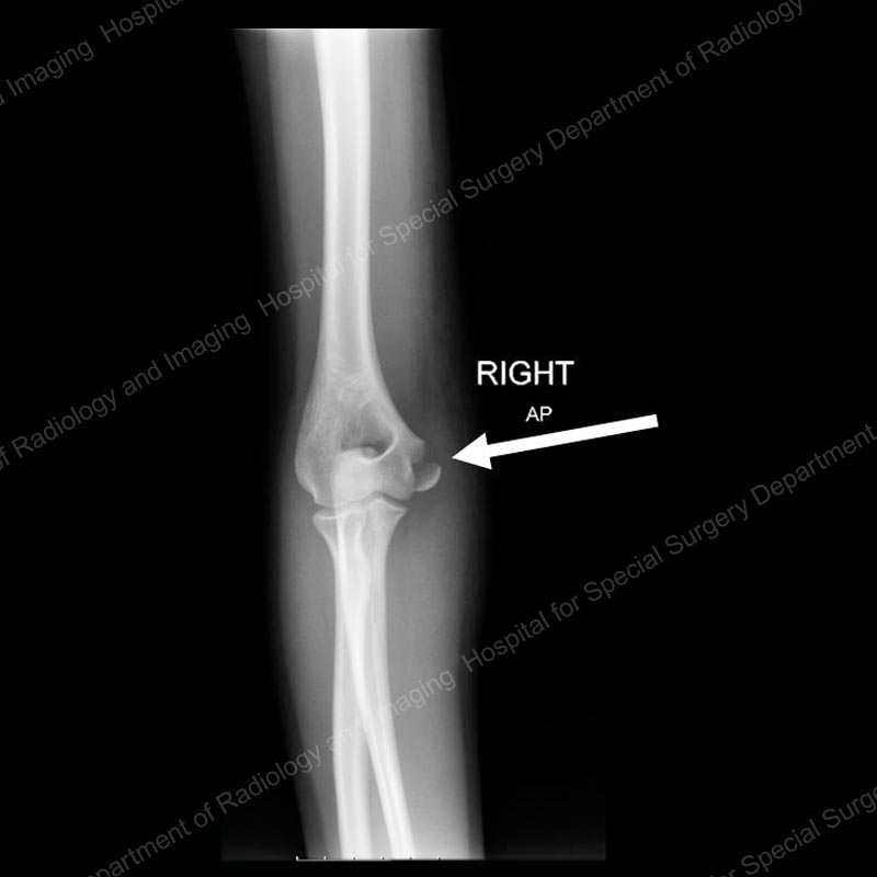
Elbow Fractures In Children An Overview Hss Edu from www.hss.edu Tendons attach muscles to bones. It develops where tendons in the forearm muscle connect to the bony part on the inside of the elbow. Tennis elbow management a multimodal management. Years at ossification (appear on xray) (1). Usually negative (evaluates more for differential diagnosis). The syndrome is also known as golfer's elbow, as it is commonly seen in. Body of humerus, surgical neck, medial supraepicondylar ridge, medial epicondyle, greater tubercle, anatomical neck. Not surprisingly, playing tennis or other racquet sports can cause this condition.
The medial epicondyle creates a prominent, blunt protuberance on the medial side of the condyle and it, is the point where the medial border of the humerus terminates by curving marginally towards the back.
Specifically in passive flexion of the elbow, it is subcutaneous and generally noticeable. Performed with the elbow extended. Most common elbow fracture in patients age <8yr. Golfer's elbow,often also called medial epicondylitis is defined as a pathologic condition that involves the pronator teres and flexor carpi radialis origins at the medial epicondyle. Incarcerated medial epicondyle fractures are commonly associated with ulnar nerve symptoms; Tendons attach muscles to bones. Tennis elbow management online course: Elbow injuries often have characteristic radiological appearances, which may only be detected by the presence of soft. Medial epicondylitis describes inflammation, pain, or tenderness in the region of the medial epicondyle of the humerus. Tenderness at the medial epicondyle. Medial epicondylitis (golfer's elbow) is a type of tendinitis that affects the inside of the elbow. Related online courses on physioplus. It develops where tendons in the forearm muscle connect to the bony part on the inside of the elbow.
Tenderness at the medial epicondyle epicondyle elbow. The top countries of suppliers are china, taiwan.


0 Comments:
Posting Komentar